Atrial Septal Defect (ASD) and Ventricular Septal Defect (VSD) are developmental abnormalities resulting in patent holes between atria and ventricles, respectively. If the defect is small, no treatment is needed besides regular check-ups. However, in cases where there is a risk of development of heart failure, a surgical occlusion as early as possible is required.
- ASD and VSD can be either treated by cardiac catheterization, or surgically corrected by direct occlusion of the defect hole with the aid of a heart–lung machine.
The ductus arteriosus is an essential blood vessel which exists between the aorta and the pulmonary artery during fetal development. The fetal vessel normally closes shortly after birth but in rare cases remains patent. This failure of closure results in excess blood flow into the lungs. The condition, which is called patent ductus arteriosus (PDA), poses a burden on the heart and progresses to heart failure. Curative treatment options include catheterization and surgery.
- PDA is the most common congenital heart disease that we see. We perform both direct surgical ligation as well as catheterization. The major advantages of direct ligation are its reliability of closure and the ability to carry out the surgery without concern for the patient weight (surgeons have experienced operating on puppies weighing between 600 and 700 grams). The benefit of catheterization lies in the minimal incision size.
PS is a condition where the pulmonary valve (or structures close to it) become narrowed, compromising blood flow to the lungs. If the narrowing is mild, no treatment or pharmacotherapy with drugs that lower heart rate may be required. With severe narrowing that imposes a heavy burden on the right heart, a minimally invasive, catheterization procedure called balloon dilation can be performed. In case of poor response to catheterization therapy, the patient will be treated by open chest surgery. The scary aspect of this disease is that symptoms are often not apparent. Thus, it is important to have a routine cardiac checkup and initiate treatment as appropriate.
- PS is treated by minimally invasive catheterization with the aid of fluoroscopy.
- In case where catheterization is contraindicated or inappropriate, pulmonary valvotomy with the aid of a heart–lung machine is performed.
AS is narrowing of the aortic valve (or structures close to it), which is essential for systemic blood supply. Pulmonary edema can occur as a complication. Like PS, AS is similar in terms of treatment strategies where mild conditions require no or only drug therapy and regular follow-ups. Severe AS cases may require a balloon dilation procedure via catheterization.
Cardiac neoplasia includes those that arise from the basal part of the heart, so-called heart base tumors, and those that arise primarily from the heart per se. The most common type of heart base tumor is called chemodectoma, in case of which pericardiotomy is the frequently performed treatment. Hemangiosarcoma often involves the right atrial appendage. Depending on the extent of infiltration, surgical resection may also be achievable.
- Heart base tumor is often not amenable to resection. However, pericardiotomy can prevent development of pericardial effusion and reportedly improve prognosis.
- Tumors arising from the right atrial appendage may be resectable. Patients with non-resectable tumors may still be surgically indicated for hemostasis in case of hemorrhage.
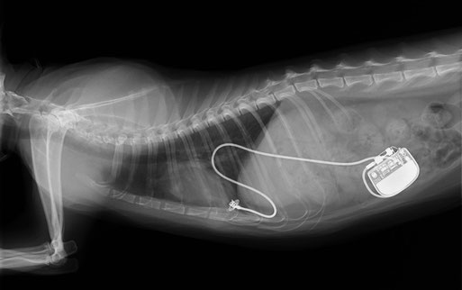
With an abnormally slow heart rate, bradyarrhythmia causes symptoms such as syncope or weakness. These can sometimes be confused with signs attributed to neurological disorders. Diagnosis is achieved by electrocardiography and Holter monitoring. This condition is generally treated by pacemaker implantation, as in humans.
- Pacemaker implantation is an effective treatment for bradyarrhythmia.
- This is the X-ray image of a cat with high-grade atrioventricular block causing recurrent syncopal episodes. Syncope completely disappeared and the cat regains its vigor.
Just like human, heart disease in animals can be broadly divided into categories: congenital, which is present at birth, and acquired, which develop after birth. In either case, proper diagnosis and treatment are always essential.